Coughing, sneezing and back pain- OH MY!

Lumbar spinal stenosis is a chronic condition characterized by a compression of the neural and/or vascular structures, which may lead to one side or two-sided pain/discomfort in the back, buttocks, thigh, calf and/or foot. Etiology: The compression arises as a result of a narrowing within the central spinal canal (as shown in the picture). This can be a result of traumas, motor vehicle accident, thickening of the osteoarthritic facet joints or bulging of degenerative discs.
Sign & Symptoms:
Tingling and numbness on both sides of low back, buttocks, thighs, calves and feet may lead to weakness with walking and prolonged standing aggravated by coughing & sneezing alleviated by sitting, prolonged rest or postural changes If you have any questions and concerns, please come in to see our physiotherapy team. We provide hands on treatment with individualized exercises program to get your back better and stronger!
If you have any questions or concerns, please do not hesitate to contact us today! 

Neck Series Part II: All about this little unique neck muscle- sternocleidomastoid (SCM)

Welcome back for another neck series.  Did everyone give the range-of-motion test a try?  Did both left, right, front, and back sides feel even and pain-free?  Hope it did!

Below is a chart to compare your ranges.  Hooray for charts!


Cervical Spine Direction Degrees of Range (Approx. degrees)
Flexion 50
Extension 60
Lateral Flexion 45
Lateral Rotation 80


Today, we will be looking at one of the most unique muscles of the neck, the sternocleidomastoid (SCM).  This muscle is located in the front of your neck on both left and right sides and controls movements associated with the neck.  It has attachments on the sternum, clavicle, and temporal bone of your skull (mastoid process), hence the name sterno-cleido-mastoid.  To locate the muscle, simply rotate your chin to one shoulder and the SCM will simply pop out and become distinct in the front of your neck.  Palpate from behind the ear, down towards the front of the neck.  Here are the anatomical all-star stats for the SCM:


Origin

(where the muscle starts)

 Manubrium (sternum)

Clavicle (collar bone)

Insertion

(where the muscle ends)

 Mastoid process (behind the ear)
Action on C-spine

(movement it produces)

Bilaterally (both sides together)

  • flexion
  • extension


Unilaterally (one side only)

  • lateral flexion (ear to shoulder)
  • lateral rotation to opposite side (chin to opposite shoulder)
Nerve Innervation

Spinal accessory nerve (XI)

Sensory supply from C2 and C3


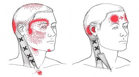
So why are we looking at this muscle?  The SCM is highly susceptible to something we call myofascial trigger points.  These points generally feel painful and very tender upon palpation.  You may even feel “knots” when palpating these muscles.  Trigger points form because the muscle is too overworked, stressed, and ultimately tight. Each muscle will have a unique referral pain away from the muscle itself.

http://www.triggerpoints.net/muscle/sternocleidomastoid


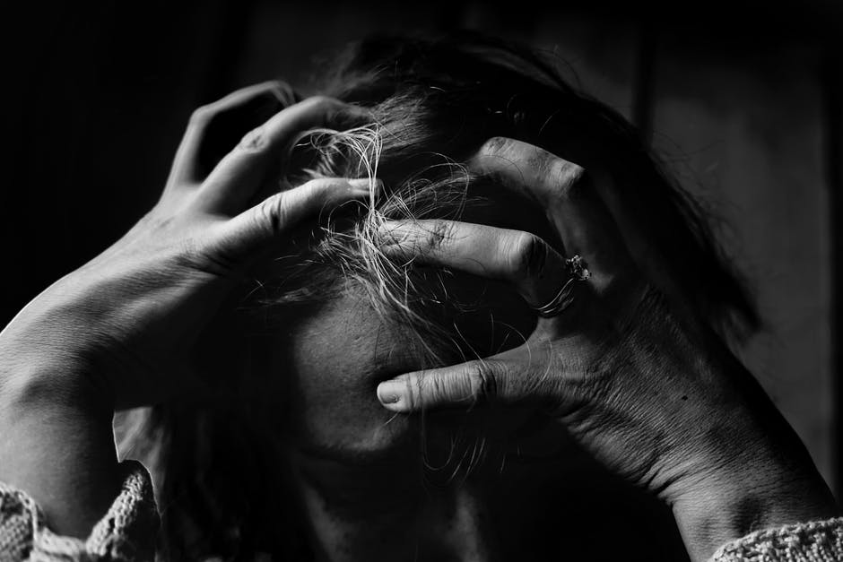
Most often, injury/dysfunction of this muscle typically comes from poor head-neck posture or from trauma such as whiplash.  Here are some common signs and symptoms you may be experiencing:


  • Neck pain
  • Headaches (occipital, temporal, frontal, or around the eyes)
  • Migraines with visual disturbances
  • Dizziness
  • Pain in upper chest


As massage therapists, when considering treatment goals, we want to make sure these trigger points are addressed and that everything is properly aligned.  Without proper alignment, the muscle will not be able to function optimally.  Here are some tips and tricks on treating your SCM:


  1. Stretch the muscle
  • Make sure your head and neck are properly aligned in a neutral position
  • Tilt your chin up slightly as if you are gazing at the stars.
  • Proceed to bring your hand over your head onto the temple
  • Bring your ear to your shoulder
  • Go slow and stretch only until you feel a nice pulling sensation along the side of the neck.  Hold the stretch for 30 to 60 seconds.
  • Make sure to avoid any unnecessary pain and keep your shoulders down.
  1. Self-release
    • Locate the muscle by first rotating the chin to one side.
  • Lightly pinch/grab the muscle belly


  • While holding onto the muscle, align yourself in a neutral position looking straight ahead.
  • Perform slow movements of ear to opposite shoulder repetitively
  • Self-releases should only be performed 20 to 30 seconds at a time with rest between sessions.

Give the stretch and self-release a try for yourself.  Hope you find some sort of relief with them!  Please stay tuned for more content including videos and more posts about the neck muscles.  Cheers!

By: Jonathan Chang, RMT, SMT(cc)


References:

http://thewellnessdigest.com/sternocleidomastoid-muscles-affects-head-eyes-sinus-ears-throat-pain-dizziness-whiplash/

https://www.physio-pedia.com/Sternocleidomastoid


Contact us today to book your massage!

Denise House Holiday Season Gift Program

Our team here at JointAction has decided to once again support the Denise House this holiday season. Founded in 1991, the Denise house provides a shelter for women and their children who suffer at the hand of domestic violence here in the Durham Region.  Below is a list of gift suggestions for Mothers and their Children, we will be collecting the items here at the clinic until December 15, 2017.


To learn more about the Denise House and their mission, click on the link below:

Piriformis Syndrome- A Pain in the Butt (LITERALLY!)

Have you experienced buttock pain? Have you being told that you have sciatica? Do you know that piriformis syndrome can also cause pain that resembles sciatica?

Piriformis muscle is located in the buttock region. When it spasms/swells/gets tightened, it can irritate the sciatic nerve that passes through the muscle. This leads to tenderness in the buttock, tingling/numbness along the back of the leg, and sometimes into the foot. You might have increased pain after prolonged sitting, reduced movement of the hip and pain with walking up stairs.

There is a quick 2 minute video that describes piriformis syndrome and sciatica. Please come in for a 15 minute free consultation if you have any questions.


By: Jie (Janet) Yang, PT.  


If you have any questions or concerns, please don’t hesitate to contact us today for you FREE 15 minute meet and greet!

Chiropractic adjustments & pregnancy: why it can be beneficial

During pregnancy there are changes in posture due to her growing belly where the abdominal muscles are stretched. This places a lot of stress into the lower back, hips, knees, ankles and feet. The average amount of weight gain during pregnancy is approximately thirty pounds. Usually as the pregnancy goes on, there is a pregnancy “penguin-like” waddle.

Physical changes are obviously visible but there are the hormonal changes which occur that you cannot see. There are hormones which increase at the end of the pregnancy which loosen up the joints in the pelvis which accommodate the uterus. The back and pelvic muscles have to work really hard to keep the spine upright and balanced.

Back pain can significantly be reduced by manual therapies including chiropractic care. The vertebrae(bones of the spine) encase the spinal cord and the proper movement of these bones can aid in the proper functioning of the nervous system. This is especially important during pregnancy as it can decrease pressure on the joints, muscles and nerves of the spine. It can help with posture and labour.

Chiropractic treatment can involve spinal adjustments, soft tissue therapy and exercise to say the least. There are various ways to adjust the spine guarantee that a pregnant patient is comfortable and safe. Using pregnancy pillows for her growing belly and tables which have a pelvic piece which drop down are helpful. There are different techniques which the chiropractor can use to adjust the spine beyond the traditional manual type of adjusting; the Activator(the main technique I use with pregnant patients usually), Sacro Occipital Technique, and Webster to name a few.

Spinal areas that I will find that are commonly restricted during pregnancy are in the upper thoracic spine from the chest increasing in size, lower thoracic spine and lumbar spine due to the expanding abdomen, the sacrum, sacroiliac joints, and  pubic bones which encase the uterus. Soft tissue trigger points are commonly found in the gluteus, piriformis, thoracic and  lumbar paraspinals, rhomboids and upper trapezius muscles.

Chiropractic alongside other conservative treatments can aid in alleviating some of the pressures built up in the body during the process. It can work optimally with other types of treatment including physiotherapy and massage as well.  Something to consider as pregnancy pre and post bring about a lot of changes to the body and should be addressed accordingly.

By: Dr. Teesha Geevarghese (B.Sc., D.C.) Chiropractor


To learn more about Chiropractic care click here!

Should you have any questions or concerns contact us for your free 15 minute meet and greet today!

My MRI shows a rotator cuff tear! What does that mean? Now what?

Rotator cuff muscles are our shoulder stabilizer muscles that also assist with shoulder movement. Our rotator cuff is made up of 4 muscles (supraspinatus, subscapularis, infraspinatus and teres minor). We often see patients with shoulder pain that come in with imaging showing a tear in one or more of the rotator cuff muscles. Some patients are able to recall a specific incident or injury that may have caused the tear and some can’t. It is important to recognize that the pain experienced may or may not be due to the rotator cuff tear as some people have rotator cuff tears and have no pain! A study found that out of 96 individuals that had no shoulder pain: magnetic resonance imaging (MRI) identified 33 of them that had rotator cuff tears including full- and partial- thickness tears1.  The study stated that the participants were able to do functional activities without any pain1. From clinical experience people with rotator cuff injuries sometimes have pain related to other muscles that have been compensating for the initial injury and are being overused. After an injury, it is natural for us to guard and protect our shoulder from painful activities but sometimes that can turn into a habit. That’s when we notice a loss of strength, range of movement, pain and various compensation strategies. Physiotherapy treatment is focused on decreasing pain, improving your range of movement and strengthening your shoulder to regain any functional limitations experienced. Remember that even patients with pain associated with a shoulder tear can return to pain-free activities. It’s important for patients to realize that they may have always had a rotator cuff tear and shoulder pain may or not be associated with the tear. Imaging is a valuable tool however we have to use and interpret findings carefully as structural changes picked up through imaging doesn’t always directly correlate with symptoms.


Reference: 1. Sher J.S, et al. Abnormal findings on magnetic resonance images of asymptomatic shoulders. The Journal of Bone and Joint Surgery. American volume. 1995 Jan; 77(1):10-5


By: Nelly Temraz, PT.


If you have any questions or concerns, please don’t hesitate to contact us today for you FREE 15 minute meet and greet!

What is Pelvic Floor Physiotherapy?- Video

“1 in 3 women suffer with incontinence but only 1 in 12 report it to their Health Care Practitioner. So bladder leaking is very common but it is never normal. Neither is painful sex, pelvic pain or urgency and frequency of urination. Pelvic health physiotherapists can help you address these problems and get you to live the life you deserve.

Please watch this short video and if you need some help please book an appointment.


By: Kate McCormick, PT

If you have any questions or concerns, please don’t hesitate to contact us today for you FREE 15 minute meet and greet!

Paediatric Milestone series: Activities in Supported Standing

Before we discuss independent standing and standing up without using support, it would be appropriate to consider simple exercises when the child is standing at the couch, ottoman, coffee table, etc. In fact, this is quite a critical period for the child to master their body in space when trying to stand and balance at their support surface.


Once the child is up and standing at their preferred support surface, placing toys or any other item of interest just out of reach on the couch is an excellent activity to practice weight shifting and challenging the boundaries of their balance while they are well supported. During a single lateral reach, the child will shift their body and take more weight on the same-sided leg and foot. This simple and subtle movement is so important because this allows the child to unweight the opposite leg and foot. With progression and strengthening, complete lifting of the opposite foot from the ground will be achieved to reach even further. Mastery of this combination of weight shifting and balancing is incredibly important because this skill is absolutely necessary for walking; weight shifting onto a single leg, lifting the opposite leg, and progressing the unweighted leg and foot forward to walk.


For continued balance and strength development, encourage rotation and reaching away from their support. Start by using a toy on the couch or table and slowly pull the toy out of reach along the surface and more towards the side of the child to encourage reaching away from their support. Continue to repeat this to both sides and within their tolerance, ensuring to give breaks as needed. Gradually increase how far the child reaches in an arc around their reaching radius until they can almost reach behind by rotating at the trunk and waist with a hand still on their support. Again, this exercise is working their ability to weight shift and balance at the same time; however, with rotating and balancing as well, we are working on trunk strengthening with movement to build strong core muscles to support the trunk and upper body when it is time for standing and walking independently.


Of course, during these activities it is important to give plenty of positive reinforcement and feedback to demonstrate the importance and excitement of standing, reaching, rotating, and moving.


We will take another detour before we get to independent standing up; we will first discuss cruising along their support on the next blog post.


By: Chris Dahiroc, PT.


Questions or concerns regarding your child’s development? Contact us for your free 15 minute meet and greet today!

Suffer from heel pain? What should I know about Plantar Fasciitis?

What is it?

Plantar fasciitis is one of the most common causes of heel pain. Plantar fascia is a thick band of tissue that originates from your heel and runs across the bottom of your foot to your toes. When you strain your plantar fascia, it becomes weak and inflamed. This type of injury will often lead to pain around your heel or in the bottom of your foot when you stand or walk, especially for the first few steps in the morning.

Common causes & Risk factors

Plantar fascia acts to support the arch of your foot. Repeated strain can cause micro-tears in the fascia. These can lead to pain and swelling.  Some of the risk factors for developing plantar fasciitis are:

  • Flat feet or high foot arches
  • Wearing shoes with poor support, such as high heels
  • Walk and stand on hard surfaces for long period of times, such as teachers and factory workers
  • Overweight
  • Middle-aged or older: most common between the ages of 40 to 60

Signs & symptoms

  • Stiffness or pain in the bottom of your heel
  • Pain travels from the bottom of the foot towards the mid-foot area
  • Pain is usually worse in the morning when you take first few steps
  • Pain tends to get better after a few steps but get worse as the day goes on
  • Pain can also be triggered by rising from sitting or standing for long periods
  • Increased pain when climbing stairs

Treatment plan

The treatment plan starts with pain relief during the early stage of plantar fasciitis. Then, it will move onto regain full movement of your foot through targeted stretches and manual techniques, and restore specific muscle strength of your foot through specific exercise. The treatment plan ends with restoring normal foot biomechanics and improving techniques to prevent recurrences to ensure a safe and functional return to work and/or sport.


By: Jie (Janet) Yang, PT.  


If you have any questions or concerns, please don’t hesitate to contact us today for you FREE 15 minute meet and greet!